What Is a 4D Ultrasound?
4D ultrasound is an advanced obstetric ultrasound technology that allows you to see the fetus as a three-dimensional moving image in real time. While 3D ultrasound provides a static three-dimensional image, 4D adds the fourth dimension of time — meaning a continuously moving 3D image, like watching a video of your baby inside the womb.
Unlike 2D ultrasound (flat black-and-white images that only doctors can interpret), 4D ultrasound allows expectant mothers and families to:
- Clearly see the baby’s face: eyes, nose, mouth, ears, cheeks — does baby look more like mom or dad?
- Observe cute gestures: thumb sucking, yawning, smiling, grimacing, kicking, waving
- Detect surface abnormalities: cleft lip/palate, limb defects (missing fingers, clubhand), abdominal wall defects
- Save memorable photos and videos — many families print 4D ultrasound images as keepsakes
With over 30 years of experience in obstetric ultrasound, BSCKI. Tran Thi Thuy Lam — Member of the Vietnam Ultrasound Association — shares detailed guidance to help mothers know when to schedule a 4D ultrasound and how to get the best images.
Comparing 2D, 3D, and 4D Ultrasound — What’s the Difference?
| Feature | 2D | 3D | 4D |
|---|---|---|---|
| Image | Flat, black and white | Static 3D (photo) | Moving 3D (video) |
| Primary purpose | Measure growth parameters, evaluate internal organs | Static surface imaging | Surface imaging + movement observation |
| Internal defect detection | Best (gold standard) | Supplementary | Supplementary |
| Surface defect detection | Limited | Good | Best |
| Easy for parents to understand | Difficult (needs doctor explanation) | Easy to see | Easiest |
| Cost | 150,000-250,000 VND | Less common | 300,000-500,000 VND |
Important: 4D ultrasound does not replace 2D ultrasound. 2D remains the standard method for measuring fetal growth parameters (BPD — biparietal diameter, FL — femur length, AC — abdominal circumference) and evaluating internal organ structures (brain, heart, lungs, kidneys). 4D ultrasound supplements with detailed surface imaging. Learn more about types of obstetric ultrasound.
Best Timing for 4D Ultrasound
Weeks 24-28: The Golden Window
This is the most ideal period for 4D ultrasound for 4 reasons:
- Baby’s facial features are fully developed — From week 24, subcutaneous fat begins forming, making baby’s face rounder and more defined. Before week 24, baby’s face is still thin with prominent cheekbones, resulting in less attractive images
- Optimal amniotic fluid volume — Amniotic fluid serves as a “window” for ultrasound waves to pass through. Weeks 24-28 have the optimal fluid level for the sharpest images
- Baby still has enough room — Baby can turn, wave, and kick, creating many beautiful angles and cute gestures
- Weeks 26-28 typically provide the sharpest images — the “golden window within the golden window”
4D Ultrasound Timing Throughout Pregnancy
| Timing | Advantages | Disadvantages | Recommendation |
|---|---|---|---|
| Weeks 16-20 | Can see baby’s entire body in one frame | Facial features unclear, skin thin and translucent | If wanting to see the whole body |
| Weeks 24-28 | Clear facial features, varied gestures, best images | — | Strongly recommended |
| Weeks 28-32 | Baby’s face is round with chubby cheeks | Less amniotic fluid, baby often covers face, less room to turn | Still good if you missed weeks 24-28 |
| Weeks 32-36 | Baby is large, clear face | Too cramped, baby often face-down against placenta | Difficult to get good images |
| Week 36+ | Baby is near birth | Very cramped, images often blurry, less movement | Not recommended |
Tip: For the best possible images, schedule your 4D ultrasound at weeks 26-28. If baby isn’t cooperative (turns away, covers face), you can come back 1-2 weeks later.
What Can 4D Ultrasound Detect?
Beyond seeing baby’s image, 4D ultrasound has important medical value:
Surface defects well-detected by 4D
- Cleft lip/palate: 4D sees this much more clearly than 2D
- Limb defects: Clubhand/foot, missing fingers, fused fingers
- Abdominal wall defects: Omphalocele, gastroschisis
- Ear defects: Microtia, anotia
- Encephalocele: Head region abnormalities
Internal organ defects (2D is still better)
- Congenital heart disease, brain defects, kidney abnormalities are best assessed with 2D
- 4D supplements 2D, especially when confirming surface defects
Note: 4D ultrasound cannot detect chromosomal abnormalities (Down, Edwards, Patau) — prenatal screening tests (Double test, Triple test, NIPT) are needed for this purpose.
Preparing for a 4D Ultrasound — Tips for the Best Images
To get the best 4D ultrasound images, mothers should prepare:
Before arriving at the clinic
- Drink orange juice or have a light snack (fruit, chocolate) 30 minutes before — sugar stimulates baby to move more, creating more cute gestures
- Don’t come when overly full — A full stomach can push the uterus up, affecting viewing angles
- Stay hydrated for 24 hours before — good amniotic fluid volume helps clearer images
- Rest well the night before — when mom is relaxed, baby is more comfortable too
At the clinic
- Wear loose clothing — Dress or elastic-waist pants, easy-to-lift top
- Come in the morning — Mom is more comfortable, baby tends to be more active
- Be patient — If baby turns away or covers their face, the doctor will ask mom to walk around, lie on her side, or wait for baby to change position. 4D ultrasound typically takes 15-30 minutes (longer than 2D)
- Bring a USB drive or phone — Ask the clinic if they save videos/images
Situations Where 4D Ultrasound Is Difficult — Solutions
4D images may not be clear in some situations:
| Situation | Cause | Solution |
|---|---|---|
| Baby facing the placenta | Unfavorable position | Mom walks 10-15 minutes, has a snack, returns |
| Low amniotic fluid (oligohydramnios) | Insufficient “window” for ultrasound waves | Drink more water, may need to reschedule |
| Overweight mother | Thick abdominal fat blocks ultrasound waves | Requires high-quality machine with good probe |
| Very large baby (late pregnancy) | Not enough room | Schedule earlier (weeks 24-28) |
| Anterior placenta | Placenta blocking baby’s face | Doctor finds alternative angles |
Note: If the first scan doesn’t capture good images, don’t be discouraged. Many clinics allow a free or discounted rescan. Ask before your ultrasound.
Is 4D Ultrasound Safe?
Completely safe. This is a question most expectant mothers worry about, so it needs clear explanation:
- Ultrasound uses sound waves — similar to those dolphins and bats use for echolocation. Not X-rays, not ionizing radiation
- WHO (World Health Organization), AIUM (American Institute of Ultrasound in Medicine), and Vietnam’s Ministry of Health all confirm obstetric ultrasound is safe
- Safe when performed multiple times during pregnancy — no accumulation, no harm
- 4D ultrasound uses the same technology as 2D, differing only in image processing
- Requirement: Must be performed by experienced specialist doctors using certified equipment
After more than 50 years of medical ultrasound use, no evidence suggests ultrasound harms mother or baby.
4D Ultrasound vs. Morphology Ultrasound — What’s the Difference?
Many mothers confuse the 4D “see baby’s face” ultrasound with the morphology scan. These serve different purposes:
| 4D “see baby’s face” | Morphology scan (weeks 20-22) | |
|---|---|---|
| Purpose | View surface images, save keepsakes | Evaluate all fetal organs |
| Timing | Weeks 24-28 (flexible) | Weeks 20-22 (required) |
| Technique | 4D (moving 3D) | Primarily 2D + supplementary 4D |
| Content | Face, limbs, gestures | Brain, heart (4 chambers), lungs, kidneys, spine, limbs |
| Required? | No (optional) | Very important — should not be skipped |
The morphology ultrasound at weeks 20-22 is an essential prenatal milestone that no mother should miss. Many clinics combine 2D morphology + 4D in a single appointment at weeks 20-22.
4D Ultrasound Costs in Lao Cai
| Facility | Estimated cost | Notes |
|---|---|---|
| Lao Cai Provincial Maternity and Children’s Hospital | 200,000 - 350,000 VND | Business hours, long wait |
| Phong Kham BS Lam | 300,000 - 500,000 VND | 7 days/week, short wait |
| Other private clinics | 250,000 - 500,000 VND | Varies by facility |
See detailed information at prenatal care costs in Lao Cai.
When to See a Doctor
During ultrasound, if the doctor detects abnormalities, they will advise on next steps. Mothers should note:
- If ultrasound reveals suspected defects, the doctor may order a repeat scan at a specialized facility or additional tests
- If oligohydramnios (low amniotic fluid) is found, more frequent monitoring is needed
- If polyhydramnios (abnormally high amniotic fluid), the cause needs investigation
- All abnormal ultrasound findings should be evaluated by a specialist doctor — don’t draw your own conclusions
Advice From a Specialist
“4D ultrasound is not just a keepsake image — it has important medical value in detecting surface defects. I recommend all expectant mothers have at least 1 4D ultrasound at weeks 26-28 for the best images, while also evaluating the baby’s surface anatomy.”
— BSCKI. Tran Thi Thuy Lam, Member of the Vietnam Ultrasound Association
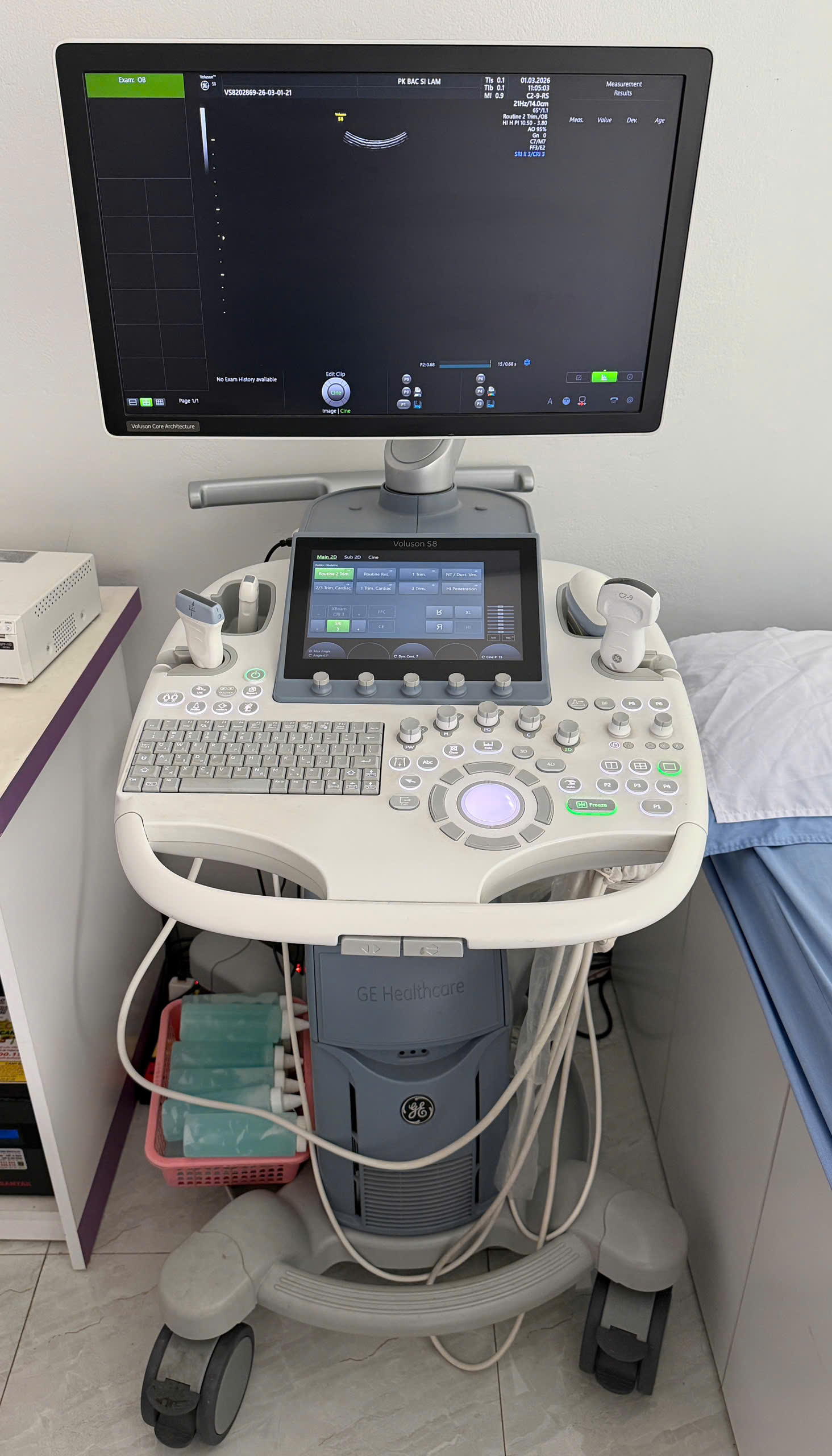
4D Ultrasound at Phong Kham San Phu Khoa Bac Sy Lam — Lao Cai
The clinic is equipped with a latest-generation high-quality ultrasound machine with dedicated probes for 4D ultrasound, providing sharp and detailed images. Ultrasounds are performed by:
- BSCKI. Pham Hong Thang — Specialist Level I in Diagnostic Imaging, Hanoi Medical University graduate — expert in fetal morphology ultrasound
- BSCKI. Tran Thi Thuy Lam — 30+ years of obstetric experience, Member of the Vietnam Ultrasound Association, formerly at Hanoi Medical University Hospital
Obstetric ultrasound services at the clinic include:
- 2D ultrasound for fetal measurements (each visit)
- 4D ultrasound for baby’s images (weeks 24-28)
- Detailed morphology ultrasound (weeks 20-22)
- Doppler ultrasound for circulation assessment (third trimester)
- Photo/video keepsakes saved for families
Book Your 4D Ultrasound
Call 0986 321 000 to schedule your 4D ultrasound.
Address: 125 Ham Nghi Street, Kim Tan, Lao Cai — Open 7 days/week, 7:00 - 19:00
A 4D ultrasound is a wonderful opportunity to meet your little one before their birthday. Book your appointment at weeks 26-28 for the best images!